Active Fibroblast Growth Factor 1, Acidic (FGF1)
酸性成纤维细胞生长因子(FGF1)活性蛋白
[ PROPERTIES ]
Source: Prokaryotic expression. Host: E. coli
Residues: Phe16~Asp155
Tags: N-terminal His-tag
Purity: >98%
Buffer Formulation: 20mM Tris, 150mM NaCl, pH8.0, containing 0.05% sarcosyl
and 5% trehalose. Applications: Cell culture; Activity Assays.
(May be suitable for use in other assays to be determined by the end user.)
Predicted isoelectric point: 7.2
Predicted Molecular Mass: 17.1kDa
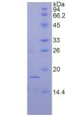
Accurate Molecular Mass: 18kDa as determined by SDS-PAGE reducing conditions.
[ USAGE ]
Reconstitute in 20mM Tris, 150mM NaCl (pH8.0) to a concentration of 0.1-1.0mg/mL. Do not vortex.
[ STORAGE AND STABILITY ]
Storage: Avoid repeated freeze/thaw cycles.
Store at 2-8℃ for one month.
Aliquot and store at -80℃ for 12 months.
Stability Test: The thermal stability is described by the loss rate. The loss ratewas determined by accelerated thermal degradation test, that is, incubate the protein at 37℃ for 48h, and no obvious degradation and precipitation were observed.The loss rate is less than 5% within the expiration date under appropriate storage condition.
[ SEQUENCE ]

[ ACTIVITY ]
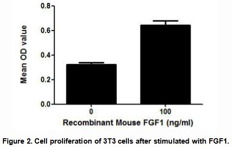
(FGF1) Fibroblast growth factor 1 belongs to the fibroblast growth factor (FGF)family. FGF1 plays an important role in the regulation of cell survival, cell division, angiogenesis, cell differentiation and cell migration. FGF1 is thought to stimulate the proliferation of 3T3 fibroblasts. Thus, a cell proliferation assay was conducted to detect the bioactivity of recombinant mouse FGF1 using 3T3 fibroblasts. Briefly, 3T3 cells were seeded into triplicate wells of 96-well plates at a density of 2,000 cells/well and allowed to attach overnight, then the medium was replaced with serum-free standard DMEM prior to the addition of various concentrations of FGF1. After incubated for 48h, cells were observed by inverted microscope and cell proliferation was measured by Cell Counting Kit-8 (CCK-8). Briefly, 10µL of CCK-8 solution was added to each well of the plate, then the absorbance at 450nm was measured using a microplate reader after incubating the plate for 1-4 hours at 37℃. Proliferation of 3T3 cells after incubation with FGF1 for 48h observed by inverted microscope was shown in Figure 1. Cell viability was assessed by CCK-8 (Cell Counting Kit-8 ) assay after incubation with recombinant FGF1 for 48h. The result was shown in Figure 2. It was obvious that FGF1 significantly increased cell viability of 3T3 cells.

[ IDENTIFICATION ]
 在线客服1号


